3d cell viability assay
The CellTiter-Glo 3D Cell Viability Assay Cat G9681 is specifically designed for determining cell viability in 3D culture models. In this assay a cell suspension is simply mixed with trypan blue and then visually examined to determine whether cells take up or exclude the dye.

3d Cell Culture Assays Biotek Applications
A Petri dish a 3D cell culture allows cells in vitro to grow in all directions similar to how they would in vivo.
. RealTime-Glo MT Cell Viability Assay. Diseases associated with CDC20 include Mosaic Variegated Aneuploidy Syndrome 1 and Mosaic Variegated Aneuploidy SyndromeAmong its related pathways are APC-Cdc20 mediated degradation of Nek2A and Cell Cycle MitoticGene Ontology GO annotations related to this gene include. Thus it functions as a regulator of a wide variety of downstream proteins including tumor suppressor proteins p53 and BRCA1 checkpoint kinase CHK2.
The ATP assay is the fastest cell viability assay to use the most sensitive and is less prone to artifacts than other viability assay methods. FLIPR Calcium Assay Kit Built as the most comprehensive portfolio of calcium reagents the kits deliver pre-optimized homogeneous fluorescence-based formulations to expedite assay development and screening of GPCR and ion channel targets. Caspase-Glo 37 Assay System.
We will continue offering the same great in vitro assay services now with even more ways to support your research. FLIPR Calcium Assay Kits provide the most comprehensive method for detecting intracellular calcium changes in a simple. The fluorescence microscope was used to observe the fluorescent signal of tumor cells.
The inverted microscope provides 125x to 60x magnification in fluorescence brightfield and color brightfield while the upright microscope enables other common applications including ELISpot slide. Based on the same reliable chemistry as the classic CellTiter-Glo Assay this new 3D assay reagent measures ATP as an indicator of viability and generates a luminescent readout that. A non-lytic fluorescent cell viability assay with multiplex.
Since LDH in the medium is the indicator of cellular cytotoxicity the assay can be used to monitor cytotoxicity from the same sample over time. To assay for viability simply add the pre-mixed alamarBlue reagent to cells in complete media no wash or cell lysis steps required incubate for one to four hours and read using either an absorbance- or fluorescence-based plate reader. Resazurin 7-Hydroxy-3H-phenoxazin-3-one 10-oxide is a phenoxazine dye that is weakly fluorescent nontoxic cell-permeable and redoxsensitive.
The CellTiter-Glo Luminescent Cell Viability Assay is a homogeneous method to determine the number of viable cells in culture based on quantitation of the ATP present which signals the presence of metabolically active cells. The kit can be used with different cell types including 3D cell models to measure cytotoxicity mediated by chemical compounds as well as cell-mediated cytotoxicity. The assay reagent penetrates large spheroids and has increased lytic capacityallowing more accurate determination of viability compared to other assay methods.
An easy-to-use plate-based luminescent assay for detecting caspase-37 activity. A homogeneous method optimized to assess viability in 3D cell culture. A bioluminescent method to kinetically monitor viability in cell culture up to 72 hours.
In the protocol presented here a viable cell will. RealTime-Glo MT Cell Viability Assay. A homogeneous method optimized to assess viability in 3D cell culture.
ホモジニアスなカスパーゼ-37 発光アッセイ G8090 G8091 G8093 G8092. Luminescent cell viability and cytotoxicity assays on the SpectraMax i3x Multi-Mode Microplate Reader Luminescent cell viability and cytotoxicity assays on the SpectraMax i3x Multi-Mode Microplate Reader. Cytation 7 Cell Imaging Multi-Mode Reader combines automated digital upright and inverted widefield microscopy with monochromator-based multi-mode microplate reading.
Unlike 2D environments eg. CellTiter-Glo 3D Cell Viability Assay. CellTiter-Fluor Cell Viability Assay.
Resazurin has a blue to purple color at pH 65 and is used in microbiological cellular and enzymatic assays because it can be irreversibly reduced to the pink-colored and highly fluorescent resorufin 7-Hydroxy-3H. Although Aire had a strong and specific effect on mTEC chromatin we were surprised to also find a variety of highly distinct mTEC subtypes characterized by lineage-defining TFs. The National Center for Advancing Translational Sciences NCATS manages the content of the Assay Guidance Manual with input from industry academia and government experts.
Figure 22 shows the results of extracting RNA from different sizes of individual 3D spheroids of HEK293 cells after measurement of cell viability using the real time assay reagent. Cell health and viability measurements provide essential insight into a broad range of biological processes and treatment responses. More than 100 authors from around the globe have contributed content to this free resource which is updated quarterly with contributions by experienced scientists from multiple disciplines working.
If necessary longer incubation times may be used for greater sensitivity without compromising cell health. Screen compounds for dermal corrosion with advanced 3D models. Diverse assay formats and reagents have been developed that measure specific aspects of cell viability corresponding to particular cellular response pathways and mechanisms of injury.
Instructions for Use of Products G7570 G7571 G7572 G7573. We own and operate 500 peer-reviewed clinical medical life sciences engineering and management journals and hosts 3000 scholarly conferences per year in the fields of clinical medical pharmaceutical life sciences business engineering and technology. The protein encoded by this gene belongs to the PI3PI4-kinase family.
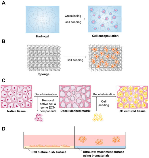
These three-dimensional cultures are usually grown in bioreactors small. CDC20 Cell Division Cycle 20 is a Protein Coding gene. A 3D cell culture is an artificially created environment in which biological cells are permitted to grow or interact with their surroundings in all three dimensions.
CellTiter-Glo 3D Cell Viability Assay. Similarly Calcein AMPI staining was used to detect the 4T1 breast cancer cell viability after IFN-γ treatment. The trypan blue staining assay allows for a direct identification and enumeration of live unstained and dead blue cells in a given population.
472 updated on 21-Aug-2022. CellTiter-Glo 3D Cell Viability Assay. Assay to assess mitochondrial toxicity.
Assays to identify cell viability. Here we used the single-cell assay for transposase-accessible chromatin with sequencing scATAC-seq to investigate mechanisms of PTA expression in individual mTECs. We are an Open Access publisher and international conference Organizer.
Gold standard method for determining cell viability based on quantitation of ATP. This protein is an important cell cycle checkpoint kinase that phosphorylates. Cited in 80 publications.
Assay Cell viabilityproliferation in 4 hr in cell samples with MTS Assay Kit ab197010. The CellTiter-Glo assay from Promega uses luciferase enzyme which requires ATP in order to generate light. Human pancreatic ductal adenocarcinoma PANC-1 and normal dermal fibroblast 3D co-cultures 5 human primary fibroblasts 6 human HuccT1 cholangiocarcinoma cells 7 human glioma cell line 8 human embryonic kidney cell line 9.
The assay reagent has increased lytic capacityallowing better penetration of large spheroid samples resulting in more accurate determination of viability compared to other assay methods. Caspase-Glo 37 Assay System.

Celltiter Glo 3d Cell Viability Assay 3d Cell Culture

Mtt Assay Results On Cell Proliferation Of 3d And 2d Cultured Cells Download Scientific Diagram

Celltiter Glo 3d Cell Viability Assay 3d Cell Culture
Ijms Free Full Text Applications Of Biomaterials In 3d Cell Culture And Contributions Of 3d Cell Culture To Drug Development And Basic Biomedical Research Html

3d Cell Culture Hts Cell Viability Complete Assay Kit K948 Biovision Inc

Get More From Your Proteomics In Gel Digestion And Efficient Peptide Extraction Are Crucial For Successful Identification Of Prote Peptides Digestion Canning
3d Cell Culture Protocols And Methods Springer Nature Experiments

Validation Of Data Obtained With Celltiter Glo 3d Cell Viability Assay Download Scientific Diagram

Pin De Mageshwari Thendral En Quick Saves En 2022

Celltiter Glo 3d Cell Viability Assay 3d Cell Culture

Celltiter Glo 3d Cell Viability Assay 3d Cell Culture

3d Cell Culture Assays Biotek Applications
Cells Free Full Text 3d In Vitro Platform For Cell And Explant Culture In Liquid Like Solids

Celltiter Glo 3d A Sensitive Accurate Viability Assay For 3d Cell Cultures

In Vitro 3d Spheroids And Microtissues Atp Based Cell Viability And Toxicity Assays Assay Guidance Manual Ncbi Bookshelf

3d Cell Culture Assays Biotek Applications

3d Culture Hts Cell Viability Assay Kit Colorimetric K2032 Biovision Inc

Celltiter Glo 3d Cell Viability Assay 3d Cell Culture
Jcm Free Full Text In Vitro Characterization Of Dental Pulp Stem Cells Cultured In Two Microsphere Forming Culture Plates Html